What is a bunionette or tailors bunion on the foot – What is a bunionette, or tailor’s bunion on the foot? This comprehensive guide dives into the world of bunionettes, exploring their definition, causes, diagnosis, treatment, prevention, and potential complications. We’ll uncover the details of this often misunderstood foot condition, offering a clear and accessible understanding.
Bunionettes, sometimes called tailor’s bunions, are bony bumps that develop on the joint at the base of the pinky toe. They’re a common foot problem that can cause pain and discomfort. This article provides a detailed look at the condition, covering everything from its causes and symptoms to the various treatment options and preventive measures you can take.
Definition and Description
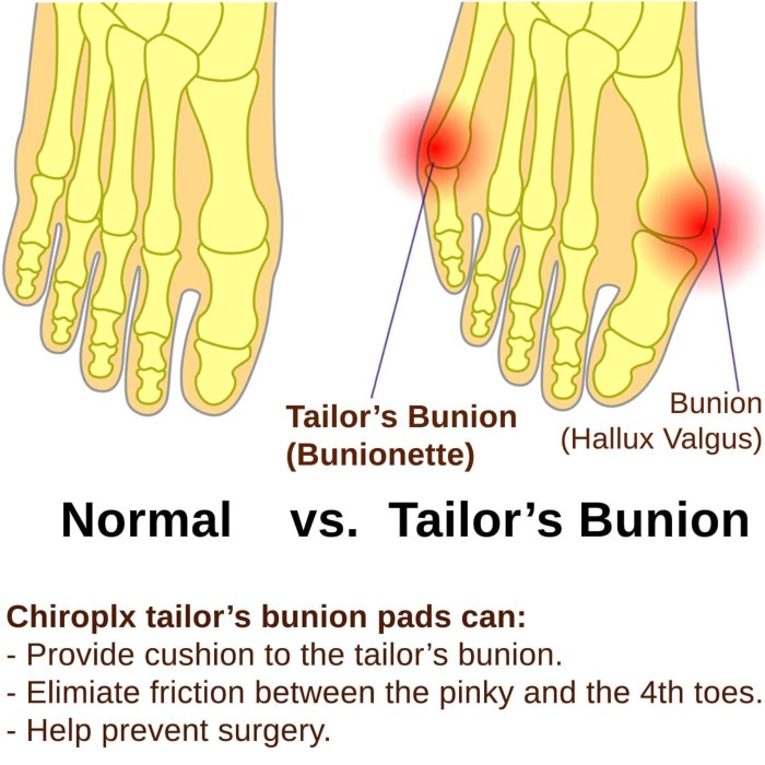
A bunionette, also known as a tailor’s bunion, is a bony bump that develops on the outside of the little toe joint. This condition causes pain and discomfort, often impacting daily activities. Understanding the specific anatomy and symptoms is key to proper diagnosis and treatment.A bunionette is a bony enlargement located on the joint at the base of the little toe.
This growth is formed by the pressure and stress exerted on the joint, causing the bone to become inflamed and enlarged. The pressure is frequently the result of repetitive stress, poor-fitting footwear, or underlying structural foot issues.
Key Differences Between Bunionette and Bunion
A bunion, on the other hand, forms on the inside of the big toe joint. While both are bony enlargements, their location and the underlying causes differ. Bunions result from the big toe deviating inward towards the other toes. Bunionettes, conversely, are characterized by a bump on the outside of the little toe.
Ever wondered what a bunionette, or tailor’s bunion, is? It’s a bony bump that forms on the little toe joint, often causing pain and discomfort. While researching foot problems, I stumbled upon an interesting article about Botox for incontinence, and how it can be used to treat certain issues. If you’re curious about whether or not botox for incontinence does it work , I recommend checking out this link.
Ultimately, though, I’m still focused on the fascinating world of foot ailments, and how a bunionette can significantly impact daily life.
Typical Symptoms of a Bunionette
Bunionettes typically manifest with a range of symptoms. The most common include pain, swelling, and tenderness around the affected area. The pain may be mild, making it tolerable for many hours or days. However, in more severe cases, the pain may be debilitating and constant.
Symptom Severity and Frequency Table
| Symptom | Severity (Mild/Moderate/Severe) | Frequency (Occasional/Frequent/Constant) |
|---|---|---|
| Pain | Mild, throbbing pain after activity; Moderate, persistent pain throughout the day; Severe, sharp, and debilitating pain | Occasional, worsening with activity; Frequent, even at rest; Constant, even with minimal movement |
| Swelling | Slight swelling around the affected area; Noticeable swelling; Significant swelling and redness | Occasional; Frequent; Constant |
| Tenderness | Slight tenderness when touched; Moderate tenderness; Severe tenderness, even with light touch | Occasional; Frequent; Constant |
| Stiffness | Minimal stiffness after prolonged rest; Moderate stiffness, hindering movement; Severe stiffness, limiting movement | Occasional; Frequent; Constant |
| Redness | Minimal redness; Noticeable redness; Significant redness, sometimes with warmth | Occasional; Frequent; Constant |
Causes and Risk Factors
Understanding the causes and risk factors of bunionettes, also known as tailor’s bunions, is crucial for prevention and management. These bony protrusions on the foot’s side can develop due to a complex interplay of genetic predisposition, footwear choices, and repetitive activities. Pinpointing these factors allows for targeted interventions and strategies to mitigate the risk of developing this condition.
Common Causes of Bunionette Development
Bunionettes, like many foot conditions, stem from a combination of factors rather than a single cause. Overuse, improper footwear, and inherited structural predispositions all contribute to the development of the bony prominence on the outside of the foot. The repetitive stress on the joint can lead to inflammation, bone spurs, and eventually the characteristic protrusion.
Risk Factors Associated with Bunionette Formation
Several factors increase the likelihood of developing a bunionette. These factors can be broadly categorized into genetic predisposition, footwear choices, and the types of activities one engages in.
Genetics and Heredity
A family history of bunionettes or other foot deformities significantly increases the risk. Genetic predisposition plays a role in the structure and function of the foot, influencing the joint’s stability and susceptibility to stress. Individuals with a family history of foot problems may have a greater likelihood of developing a bunionette, highlighting the importance of genetic factors in foot health.
Impact of Footwear Types
High heels, narrow-toe shoes, and shoes with inadequate arch support can significantly contribute to bunionette development. The pressure exerted on the outer foot by ill-fitting or constrictive footwear can irritate the joint, leading to inflammation and the formation of a bunionette. Pointed-toe shoes, for example, force the toes into a cramped position, increasing the stress on the little toe joint and surrounding structures.
Impact of Different Activities
Repetitive activities that place significant stress on the feet, such as running, dancing, or certain types of work involving prolonged standing or walking, can elevate the risk. The constant impact and pressure on the joints can contribute to the development of bunionettes over time. Activities like ballet dancing, for instance, often involve extensive, repetitive movements and foot positions that put high stress on the lateral foot joints.
Comparison of Causes and Risk Factors
| Category | Description | Examples |
|---|---|---|
| Genetics | Family history of foot deformities, inherited structural predisposition. | A parent with bunionettes, a history of flat feet in the family. |
| Footwear | Ill-fitting shoes, high heels, narrow-toe shoes, shoes lacking arch support. | Constrictive shoes, high heels, pointed-toe pumps. |
| Activities | Repetitive activities with high impact or pressure on the feet. | Running, dancing, prolonged standing, certain work requiring continuous foot movement. |
Diagnosis and Evaluation
Figuring out if you have a bunionette, also known as a tailor’s bunion, involves a systematic approach that combines a physical examination with diagnostic tools. Accurate diagnosis is crucial for developing an effective treatment plan. A proper evaluation considers the patient’s symptoms, medical history, and physical findings to arrive at an accurate diagnosis.
Typical Methods for Diagnosing a Bunionette
A thorough assessment begins with a detailed patient history. This includes inquiring about the onset, progression, and location of pain, as well as any associated symptoms like swelling or redness. The history also includes any pre-existing conditions, previous injuries, and the patient’s lifestyle, including activities that might aggravate the condition. Understanding the patient’s perspective and personal experiences is paramount in reaching an accurate diagnosis.
Role of Physical Examinations in Diagnosing Bunionettes
Physical examinations play a critical role in identifying bunionettes. The examiner will carefully inspect the affected foot and ankle, paying close attention to the positioning of the fifth metatarsal bone, the presence of any bony prominences, and the degree of any associated swelling or inflammation. Palpation, or feeling with the hands, is used to determine the tenderness and pain associated with the affected area.
This examination helps to determine the severity of the condition and any accompanying musculoskeletal issues.
Diagnostic Tools and Techniques Used by Medical Professionals
Medical professionals utilize a range of tools and techniques to confirm the diagnosis. These tools often include specialized instruments for precise measurements and assessments, ensuring a thorough evaluation of the affected area. X-rays are essential for visualizing the bone structure and identifying the degree of deviation in the fifth metatarsal bone.
A bunionette, or tailor’s bunion, is that painful bump on the pinky toe joint. It’s often caused by ill-fitting shoes or certain activities. While managing foot pain is important, it’s equally vital to focus on overall well-being, like learning to live well with allergic asthma, which can significantly impact quality of life. Thankfully, there are excellent resources available, such as strategies for living well with allergic asthma , that can help improve your daily life.
Ultimately, understanding the causes and management of bunionettes is key to preventing discomfort and enjoying comfortable feet.
Structured Overview of the Diagnostic Process
The diagnostic process for a bunionette typically follows a structured approach. First, a comprehensive patient history is taken to understand the patient’s symptoms and medical background. Following this, a physical examination is performed to assess the affected area and identify any abnormalities. If necessary, diagnostic imaging, such as X-rays, may be ordered to confirm the diagnosis and evaluate the extent of the condition.
This structured approach ensures a thorough and accurate evaluation of the bunionette.
So, you’ve got a bunionette, or tailor’s bunion, on your foot? It’s that bump on the joint at the base of your pinky toe. While we’re on the subject of foot ailments, have you ever wondered if vitamin D supplements might affect your skin tone? For more info on that, check out this article on does vitamin d supplement make skin darker.
It’s a common question, and understanding the potential effects is important. Regardless, bunionettes can be quite uncomfortable, impacting your everyday life. It’s definitely worth exploring treatment options if they’re causing you problems.
How X-rays and Other Imaging Techniques are Used to Diagnose a Bunionette
X-rays are a cornerstone of the diagnostic process for bunionettes. They provide detailed images of the bones, allowing medical professionals to visualize the alignment of the fifth metatarsal bone and the degree of its deviation. X-rays help to determine the severity of the condition and identify any underlying bone abnormalities. In some cases, other imaging techniques, such as CT scans or MRIs, may be employed to gain a more comprehensive understanding of the condition, although X-rays are usually sufficient for bunionette diagnosis.
A comparison of the affected foot with the unaffected foot helps in identifying the degree of deviation.
Table of Diagnostic Procedures and Their Applications
| Diagnostic Procedure | Application |
|---|---|
| Patient History | Understanding the patient’s symptoms, medical history, and lifestyle factors. |
| Physical Examination | Assessing the affected area, identifying bony prominences, and evaluating associated swelling or inflammation. |
| X-rays | Visualizing the alignment of the fifth metatarsal bone and the degree of deviation. |
| CT Scan (occasionally) | Providing a more detailed view of the bone structure, especially if complex bone issues are suspected. |
| MRI (rarely) | Evaluating soft tissue structures around the affected area, such as ligaments and tendons, if soft tissue involvement is suspected. |
Treatment Options and Management

Bunionettes, or tailor’s bunions, can significantly impact a person’s quality of life. Fortunately, several treatment options are available, ranging from conservative measures to surgical interventions. Understanding these options allows individuals to make informed decisions regarding their care and achieve optimal outcomes.
Conservative Approaches
Conservative treatments aim to alleviate symptoms and prevent further progression of the bunionette without surgery. These methods are often the first line of defense for managing the condition.
- Padding and Cushioning: Using specialized padding or heel cups can help reduce pressure on the affected area and alleviate pain. Custom-molded orthotics can also provide additional support and cushioning. This simple approach can significantly improve comfort and reduce pain for many patients, particularly in the early stages of the condition.
- Proper Footwear: Choosing footwear with a wide toe box and adequate support is crucial. Avoid shoes with narrow toe boxes or high heels that exacerbate pressure on the bunionette. Wearing shoes that fit properly is essential in managing the symptoms and preventing further irritation.
- Ice and Heat Therapy: Applying ice packs for 15-20 minutes at a time can help reduce inflammation and pain. Heat therapy, such as warm compresses, can also be beneficial for improving blood flow and relaxing the muscles surrounding the bunionette.
- Pain Medications: Over-the-counter pain relievers, such as ibuprofen or naproxen, can help manage pain and inflammation. In some cases, a doctor may prescribe stronger pain medications, especially if the pain is severe or persistent. It is important to follow the dosage instructions and consult a healthcare professional before using any medication.
Orthotics and Supportive Footwear
Custom orthotics and supportive footwear play a vital role in managing bunionette symptoms. They provide targeted support and help to redistribute pressure, thus reducing pain and inflammation.
- Custom Orthotics: These specialized inserts are designed to fit the individual’s foot and provide tailored support to the bunionette area. They can help to control the foot’s alignment, reduce pressure, and improve comfort. Custom orthotics often prove to be highly effective in managing bunionette symptoms.
- Supportive Footwear: Selecting shoes with adequate arch support and a wide toe box is crucial. This type of footwear helps to cushion the foot, reduce pressure on the bunionette, and maintain proper foot alignment. Supportive footwear can contribute significantly to pain relief and overall comfort.
Surgical Options
Surgical intervention is typically reserved for cases where conservative treatments have failed to provide adequate relief or where the bunionette significantly impacts a person’s ability to perform daily activities.
- Excision and Soft Tissue Procedures: Surgical procedures often involve removing the inflamed or bony prominence, as well as correcting any underlying soft tissue abnormalities. The surgeon meticulously removes the problematic bone and soft tissues to alleviate pressure and pain. This surgical approach has a high success rate in alleviating bunionette symptoms and restoring foot function.
- Osteotomy Procedures: In some cases, osteotomy, a procedure that involves repositioning bones, may be considered. This technique involves realigning the affected bone to correct the underlying structural issue, thus reducing the pressure and improving the foot’s overall function. Osteotomy procedures are usually more complex and require a longer recovery period.
Comparison of Treatment Options
| Treatment Option | Efficacy | Recovery Time | Advantages | Disadvantages |
|---|---|---|---|---|
| Conservative Treatments | Variable; often effective for mild cases | Short to moderate | Non-invasive, less expensive | Limited effectiveness for severe cases, potential for recurrence |
| Orthotics and Supportive Footwear | Moderate to high | Variable; usually gradual improvement | Non-invasive, can be used alongside other treatments | May not be suitable for all individuals, potential for discomfort during adjustment |
| Surgical Options | High | Moderate to long (several weeks to months) | Permanent solution for severe cases, often restoring full function | Involves risk of complications, longer recovery period, potential for scarring |
Prevention and Self-Care Strategies: What Is A Bunionette Or Tailors Bunion On The Foot

Bunionettes, also known as tailor’s bunions, can be a persistent issue if not managed proactively. Taking preventative steps and practicing effective self-care can significantly reduce discomfort and the likelihood of the condition worsening. Understanding how to select appropriate footwear and incorporating exercises into your routine are key components in managing this condition.Proper footwear plays a crucial role in preventing and managing bunionette pain.
Choosing shoes that offer adequate support and room for your toes is essential. Avoid tight-fitting or pointed-toe shoes that can exacerbate the condition. Selecting shoes with a wide toe box and good arch support can help maintain proper foot alignment.
Preventive Measures
Maintaining good foot health is crucial in preventing bunionettes. This includes paying close attention to the types of shoes you wear, avoiding prolonged standing or walking, and maintaining a healthy weight. By proactively addressing these factors, you can significantly reduce your risk of developing this condition.
- Appropriate Footwear: Prioritize shoes with a wide toe box, avoiding pointed or narrow styles. Look for shoes with good arch support and cushioning to reduce stress on the foot.
- Weight Management: Maintaining a healthy weight can lessen the stress on your feet and joints, thus reducing the risk of developing bunionettes.
- Avoiding Prolonged Standing or Walking: If your job or activities involve prolonged standing or walking, take breaks to rest your feet and stretch. Elevate your feet periodically.
- Regular Foot Exams: Schedule regular checkups with a podiatrist to monitor your foot health and address any developing issues early.
- Proper Posture: Maintaining good posture when standing and walking can help distribute weight evenly throughout your feet, reducing stress on specific areas.
Footwear Selection
Choosing the right footwear is paramount in preventing and managing bunionettes. Ill-fitting shoes contribute significantly to the development and worsening of the condition. Shoes that are too tight or have narrow toe boxes put excessive pressure on the affected area, exacerbating discomfort and potentially leading to further complications.
- Avoid High Heels: High heels tend to place increased pressure on the ball of the foot and can contribute to bunionette formation.
- Consider Wide Toe Boxes: Look for shoes with ample room for your toes to spread naturally. A wide toe box helps prevent crowding and pressure on the affected area.
- Check for Arch Support: Shoes with good arch support help maintain the natural alignment of the foot, reducing strain on the joints and preventing bunionettes from worsening.
- Choose Cushioned Insoles: Cushioned insoles can provide additional comfort and support, particularly when engaging in activities that place stress on your feet.
Self-Care Strategies, What is a bunionette or tailors bunion on the foot
Implementing effective self-care strategies can significantly alleviate bunionette symptoms and improve overall foot comfort. Simple measures like icing, padding, and stretching can make a substantial difference in managing discomfort.
- Ice Application: Applying ice packs to the affected area for 15-20 minutes at a time, several times a day, can help reduce swelling and inflammation.
- Padding: Using soft padding or bunionette shields inside your shoes can provide cushioning and reduce pressure on the affected area.
- Stretching Exercises: Regular stretching exercises can help maintain flexibility and reduce tightness in the affected area.
- Over-the-Counter Pain Relievers: Using over-the-counter pain relievers like ibuprofen or naproxen can help manage pain and inflammation.
Managing Pain and Discomfort
Managing pain and discomfort associated with bunionettes is crucial for maintaining comfort and mobility. Addressing the cause of the pain and implementing appropriate strategies can significantly improve your quality of life.
- Rest and Elevation: Resting the affected foot and elevating it when possible can help reduce swelling and inflammation.
- Use of Orthotics: Custom or over-the-counter orthotics can provide added support and cushioning, reducing stress on the affected area.
- Consider Shoe Inserts: Shoe inserts or padding can help alleviate pressure on the bunionette and improve comfort.
Exercise and Stretching
Regular exercise and stretching are essential for maintaining foot health and flexibility. Incorporating these into your routine can help prevent bunionettes and manage existing symptoms. Strengthening the muscles in your feet and ankles can provide better support and reduce the risk of injury.
- Toe Stretches: Regular toe stretches can help maintain flexibility and reduce tightness in the toes and surrounding areas.
- Foot Strengthening Exercises: Incorporate exercises that target the muscles in your feet to improve support and stability.
- Gentle Ankle Rotations: Gentle ankle rotations can help maintain flexibility and range of motion in the ankle joint.
Complications and Associated Conditions
Bunions, particularly bunionettes, aren’t just cosmetic concerns. They can lead to a cascade of problems if left untreated. Understanding the potential complications is crucial for proactive management and preventing further discomfort and disability. The progression of bunionette issues can significantly impact a person’s quality of life, affecting daily activities and overall foot health.
Potential Complications of Bunionette Conditions
Bunionettes, also known as tailor’s bunions, can cause a range of issues beyond the immediate discomfort of the bump on the foot. These complications often arise from the structural changes in the foot and the resulting pressure and irritation.
Connection to Other Foot Conditions
Bunionettes frequently coexist with other foot problems. Overpronation, a common cause of foot pain, can exacerbate bunionette development. Likewise, conditions like hammertoe or hallux valgus (big toe bunion) can emerge or worsen as a result of the biomechanical changes induced by a bunionette. This interconnectedness highlights the importance of comprehensive foot care and addressing underlying biomechanical issues.
Impact on Overall Foot Health
The persistent pressure and irritation associated with bunionettes can lead to chronic inflammation, pain, and structural damage. This can progressively weaken the supportive tissues in the foot, potentially leading to arthritis or other degenerative joint conditions. Regular monitoring and appropriate treatment are essential to mitigate these risks.
Progression to Further Problems
Left untreated, bunionettes can cause considerable discomfort and impact mobility. As the condition worsens, the deformity can lead to more significant structural changes, resulting in an increased likelihood of other foot problems like calluses, corns, and bursitis. The pain can become more intense and frequent, interfering with daily activities. In severe cases, the structural damage may necessitate surgical intervention.
Impact on Walking and Daily Activities
The pain and discomfort associated with bunionettes can make walking and performing daily activities challenging. The altered foot structure can make it difficult to maintain balance, leading to falls or accidents. This can severely impact an individual’s ability to participate in work, sports, and social activities. Proper footwear and supportive devices can help alleviate these issues and maintain mobility.
Categorization of Potential Complications
- Mechanical Issues: Bunionettes alter the biomechanics of the foot, increasing the risk of other foot conditions such as hammertoe, hallux valgus, and overpronation. This altered biomechanics can lead to issues like instability, pain, and reduced mobility. These issues, if not addressed, can lead to more severe complications.
- Pain and Inflammation: Persistent pressure and irritation from the bunionette can lead to chronic pain, swelling, and inflammation. This inflammation can damage the surrounding tissues, potentially leading to conditions like bursitis or arthritis. The pain can significantly limit an individual’s ability to engage in daily activities.
- Structural Damage: Over time, the misalignment of bones and tissues caused by bunionettes can result in permanent structural changes to the foot. This damage can compromise the foot’s support system, leading to a cascade of additional problems. Examples include the breakdown of cartilage and tendons, resulting in chronic pain and reduced mobility.
- Functional Limitations: The pain and structural changes associated with bunionettes can severely restrict an individual’s ability to perform daily activities. This can impact their ability to walk, stand, and participate in various physical activities, leading to a reduced quality of life. The inability to wear suitable footwear due to the discomfort is also a common functional limitation.
Illustrations and Visual Aids
Understanding bunionettes, also known as tailor’s bunions, requires a visual understanding of their location, development, and impact on the foot. Visual aids help clarify the complexities of this condition and guide individuals towards a better comprehension of their potential treatment options. Illustrations and diagrams offer a tangible representation of the anatomical structures involved, providing a crucial perspective for patients and healthcare professionals alike.
Detailed Visual Representation of a Bunionette
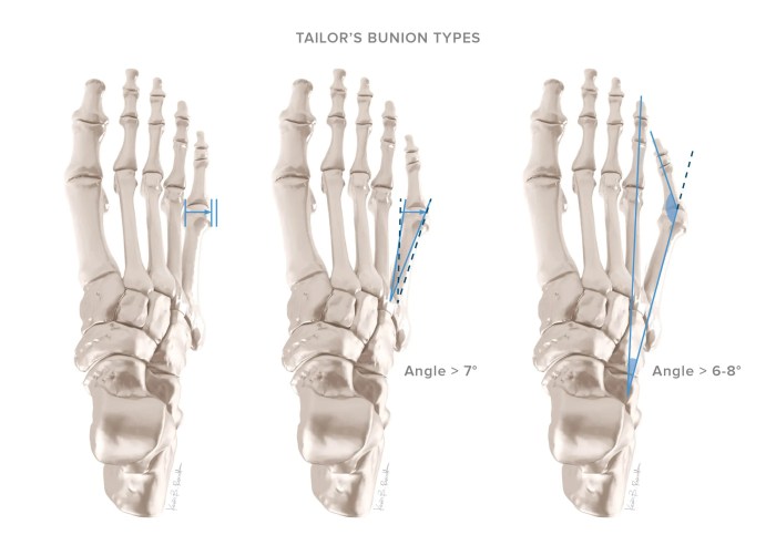
A bunionette is characterized by a bony protrusion on the outside of the little toe joint. Visual representations from various angles are essential for understanding the condition’s appearance. A front-view illustration would showcase the prominent bump, while a side-view would highlight the joint’s misalignment. A dorsal (top) view would show the affected area’s position relative to the other toes and the overall foot structure.
These perspectives collectively demonstrate the characteristic deviation of the fifth metatarsal bone, creating the bunionette.
Foot Structure with Bunionette Location
A graphic representation of the foot’s anatomy, emphasizing the metatarsals, should clearly indicate the location of the bunionette. The fifth metatarsal bone would be highlighted as the site of the bony enlargement. The diagram should also depict the surrounding soft tissues, including tendons and ligaments, to show the intricate interplay of these structures in the affected area. This visual representation is crucial for comprehending the anatomical context of the bunionette’s development.
Impact of Bunionette on the Joint
A diagram illustrating the impact of a bunionette on the joint should show the misalignment of the fifth metatarsophalangeal joint. The diagram should depict the abnormal angle of the fifth metatarsal bone, highlighting how it presses against the surrounding tissues. This visualization should also show how the misalignment can lead to pressure and irritation within the joint, causing pain and discomfort.
The illustration should clearly demonstrate the altered biomechanics within the joint.
Treatment Options Visualized
Visual representations of treatment options, such as orthotics and surgical interventions, should be presented. Illustrations of customized orthotics that specifically address the bunionette’s impact on the foot’s biomechanics can be included. Images showcasing different surgical procedures, such as bone repositioning or joint fusion, can provide a clear understanding of the surgical approaches. Visual representations can highlight the potential benefits and risks associated with each treatment option.
Anatomical Diagram of the Affected Area
A detailed anatomical diagram should focus on the fifth metatarsophalangeal joint. The diagram should clearly label the bones, tendons, ligaments, and surrounding soft tissues. It should depict the specific structures that are affected by the bunionette, including the joint capsule, and the impact of the bony enlargement on the surrounding soft tissues. This diagram would be crucial for understanding the intricate anatomy of the area.
Bunionette Stages Illustrated
| Stage | Description | Illustration |
|---|---|---|
| Mild | A small bump on the outside of the little toe joint, minimal pain, and no significant joint misalignment. | [Imagine a subtle bump on the foot’s outside, near the little toe joint. The bump is not overly prominent.] |
| Moderate | A more pronounced bony protrusion, increasing pain and some noticeable joint misalignment. | [Imagine a slightly larger bump, with a more noticeable angle in the affected joint. The surrounding skin may show slight redness or inflammation.] |
| Severe | A significant bony enlargement, substantial pain, noticeable joint deformity, and possible difficulty with everyday activities. | [Imagine a large, prominent bump on the outside of the little toe, with a pronounced misalignment of the joint. The surrounding tissues may show increased inflammation and redness.] |
The table illustrates the progression of bunionette severity, from mild to moderate to severe. Visual representations aid in understanding the visible changes associated with each stage. This aids in diagnosis and helps patients understand the expected progression of the condition.
Closing Notes
In conclusion, bunionettes, while often a source of discomfort, are manageable with the right approach. Understanding the causes, symptoms, and available treatments empowers you to take control of your foot health. By prioritizing proper footwear, maintaining a healthy weight, and seeking professional advice when needed, you can significantly reduce the risk of developing or exacerbating this condition. This guide has provided a thorough overview, enabling you to navigate the world of bunionettes with greater confidence and knowledge.


