Peroneus longus muscle anatomy explores the intricate details of this crucial lower limb muscle. We’ll journey through its location, relationships with surrounding structures, and its vital role in foot movement and stability. Delving into its microscopic structure, nerve supply, and clinical significance, we’ll gain a comprehensive understanding of this often-overlooked component of the human body.
This detailed exploration of the peroneus longus muscle anatomy covers everything from its origin and insertion points to its functions in ankle and foot movements. We’ll also look at potential injuries, variations, and how it appears in various imaging modalities.
Overview of Peroneus Longus Muscle

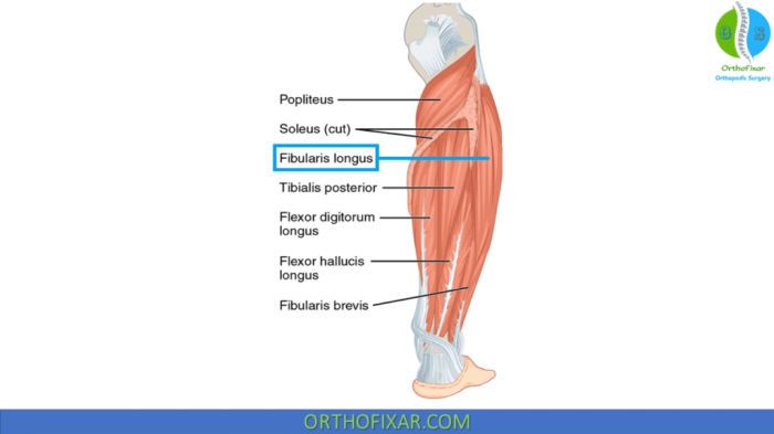
The peroneus longus muscle, a crucial component of the lower leg’s musculature, plays a vital role in foot movement and stability. Understanding its precise location, attachments, and functions is essential for comprehending its contribution to overall lower limb biomechanics. This discussion delves into the anatomical specifics of the peroneus longus, highlighting its relationship to surrounding structures and its critical functions.The peroneus longus muscle resides in the lateral compartment of the lower leg, situated superficially alongside the peroneus brevis muscle.
Its position, close to the fibula and lateral malleolus, allows it to influence foot movements effectively. This strategic placement also necessitates an understanding of its interaction with adjacent structures to fully grasp its function.
Location and Relationships
The peroneus longus muscle occupies a significant portion of the lateral compartment of the lower leg, often extending distally from the head of the fibula to the foot. Its superficial location allows for easy palpation in the region, making it readily identifiable during physical examinations. The peroneus longus lies in close proximity to the peroneus brevis muscle, which contributes to its overall role in foot movement.
Importantly, its relationship to the lateral malleolus of the ankle bone, and the tendons passing beneath it, is crucial for its function.
Origin
The peroneus longus muscle originates from the head and upper two-thirds of the lateral surface of the fibula. Precisely, its origin encompasses the anterior surface of the fibula, including the head, the upper two-thirds of the lateral surface, and the adjacent intermuscular septa. This extensive origin area provides a substantial anchor point for the muscle’s action.
Insertion
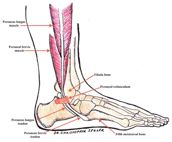
The peroneus longus muscle inserts into the medial cuneiform bone and the base of the first metatarsal bone of the foot. This insertion point is crucial for its primary function, which involves plantarflexion and eversion of the foot. The specific bony landmarks of the insertion are the medial cuneiform bone and the base of the first metatarsal bone, where the tendon attaches, enabling the muscle to exert its influence on the foot’s position and movement.
Functions
| Function | Description | Importance | Related Actions |
|---|---|---|---|
| Plantarflexion | Movement of the foot downward at the ankle joint. | Essential for maintaining posture and propelling the body forward during walking and running. | Walking, running, jumping |
| Eversion | Turning the sole of the foot outward. | Crucial for maintaining foot stability during weight-bearing activities. | Walking, standing, maintaining balance |
| Foot Arch Support | Assists in maintaining the longitudinal arch of the foot. | Prevents collapse of the arch, reducing strain on other foot structures. | Standing, walking, jumping |
| Tibiotalar joint stability | Contributes to the stability of the ankle joint. | Helps prevent excessive inversion and eversion of the ankle, reducing the risk of injuries. | Walking, running, any activity requiring ankle support. |
Muscle Structure and Histology
The peroneus longus muscle, a crucial component of the lower limb’s dynamic movement, possesses a complex internal structure that directly influences its function. Understanding this microscopic architecture is key to appreciating the muscle’s role in foot and ankle stability and mobility. This intricate arrangement of muscle fibers, fascicles, and connective tissues allows for precise control and force generation during various activities.The microscopic organization of the peroneus longus muscle, including the types of muscle fibers and their arrangement, significantly impacts its contractile properties and overall performance.
This structural layout, in conjunction with the surrounding connective tissues, ensures both efficient force transmission and optimal protection.
Microscopic Structure of Muscle Fibers
The peroneus longus, like all skeletal muscles, is composed of numerous muscle fibers, each a single cylindrical cell. These fibers are multinucleated, with nuclei located peripherally. Within each fiber, numerous myofibrils are organized into repeating units called sarcomeres, the fundamental contractile units of muscle. The arrangement of these sarcomeres creates the characteristic striated appearance of skeletal muscle tissue under a microscope.The peroneus longus contains a mixture of different fiber types, each with distinct characteristics that influence its function.
These fiber types are classified based on their contractile properties, primarily speed of contraction and energy source. This diversity allows the muscle to adapt to varying demands during movement.
Fiber Types and Their Characteristics
| Fiber Type | Contraction Speed | Energy Source | Fatigue Resistance |
|---|---|---|---|
| Type I (Slow-twitch) | Slow | Aerobic (oxidative) | High |
| Type IIa (Fast-oxidative-glycolytic) | Fast | Aerobic (oxidative) and anaerobic (glycolytic) | Intermediate |
| Type IIx (Fast-glycolytic) | Fast | Anaerobic (glycolytic) | Low |
Arrangement of Muscle Fascicles
The peroneus longus muscle’s fascicles are arranged in a somewhat oblique fashion, with a slightly more parallel orientation near the muscle’s insertion point. This arrangement is critical to the muscle’s function. The oblique orientation allows for a wider range of motion and a greater ability to generate force, particularly during eversion of the foot. The more parallel arrangement near the insertion point optimizes the muscle’s ability to exert force at the ankle and foot.
Connective Tissue Components, Peroneus longus muscle anatomy
Surrounding the muscle fibers are various connective tissue layers that contribute to the muscle’s structure and function. These layers provide support, protection, and a pathway for blood vessels and nerves. The endomysium surrounds individual muscle fibers, the perimysium encloses bundles of fibers (fascicles), and the epimysium forms the outermost layer of the muscle. These layers work together to transmit force generated by the muscle fibers to the surrounding tissues and bones.
The peroneus longus muscle, crucial for foot and ankle movement, is a fascinating part of the human anatomy. Understanding its intricate structure helps us appreciate the complexity of the body. While not directly related, learning about the symptoms of sexually transmitted infections, like gonorrhea, can be important for overall health. For a detailed guide on what gonorrhea looks like, check out this helpful resource: what does gonorrhea look like.
Ultimately, a thorough understanding of the peroneus longus muscle’s role in our lower body mechanics is key.
| Connective Tissue | Description | Function | Location |
|---|---|---|---|
| Endomysium | Delicate layer of connective tissue surrounding individual muscle fibers. | Provides support and insulation to individual muscle fibers. | Surrounding individual muscle fibers |
| Perimysium | Connective tissue that bundles muscle fibers into fascicles. | Provides support and structure to the fascicles. | Surrounding bundles of muscle fibers (fascicles) |
| Epimysium | Dense, fibrous connective tissue surrounding the entire muscle. | Protects the muscle and provides a pathway for blood vessels and nerves. | Outermost layer of the muscle |
| Fascia | Sheet-like connective tissue surrounding the muscle, separating it from adjacent structures. | Provides support, protection, and compartmentalization of muscles. | Surrounds the entire muscle, separating it from adjacent structures |
Nerve Supply and Blood Supply
The peroneus longus muscle, crucial for foot and ankle movements, relies on a complex interplay of nerves and blood vessels for its function. Understanding these pathways is essential for comprehending its role in the body and recognizing potential issues related to its performance. Proper nerve and blood supply ensure adequate oxygen and nutrient delivery, and facilitate proper signal transmission for muscle contraction.The peroneus longus muscle, like all muscles, receives both nerve impulses and blood supply, vital for its proper function.
Nerve supply dictates when and how the muscle contracts, while blood vessels deliver the necessary oxygen and nutrients to fuel these contractions. Disruptions to either system can lead to muscle weakness, pain, and potentially other complications.
Nerve Supply
The peroneus longus muscle receives its nerve supply from the common peroneal nerve (also known as the common fibular nerve). This nerve arises from the sciatic nerve and branches into two major divisions: the superficial peroneal nerve and the deep peroneal nerve. The deep peroneal nerve provides the innervation for the peroneus longus muscle. This complex pathway ensures proper signal transmission for controlled muscle actions.
Blood Supply
The peroneus longus muscle, like all tissues in the body, receives blood supply from a network of arteries. The blood vessels supplying the peroneus longus muscle are primarily branches of the anterior tibial artery and the peroneal artery. These arteries deliver oxygenated blood to the muscle fibers, enabling them to perform their functions. The venous system, a network of veins, carries deoxygenated blood back to the heart, completing the circulatory loop.
This intricate system of arteries and veins ensures a continuous flow of nutrients and oxygen to the muscle.
Nerve Supply Details
The following table Artikels the specific nerve branches responsible for innervating the peroneus longus muscle.
| Nerve | Origin | Branch | Target Muscle |
|---|---|---|---|
| Common Peroneal Nerve | Sciatic Nerve | Deep Peroneal Nerve | Peroneus Longus |
Blood Supply Comparison
The following table contrasts the arterial and venous systems that provide blood supply to the peroneus longus muscle. The arterial system brings oxygenated blood, while the venous system carries away deoxygenated blood.
| System | Artery(ies) | Venous Drainage | Description |
|---|---|---|---|
| Arterial | Anterior Tibial Artery, Peroneal Artery | Veins accompanying the arteries | Deliver oxygenated blood to the muscle tissue |
| Venous | N/A | Veins accompanying the arteries | Return deoxygenated blood to the heart |
Actions and Movements
The peroneus longus muscle, a key player in foot and ankle mechanics, plays a crucial role in maintaining balance and enabling various movements. Understanding its actions is vital for comprehending its significance in overall lower limb function. Its precise contributions to foot stability, ankle movement, and the intricate interplay between plantarflexion, eversion, and other motions are important aspects of its function.The peroneus longus muscle’s primary actions are focused on foot eversion and plantarflexion.
Its location and attachments, combined with its leverage, make it a primary contributor to these movements. Understanding these actions allows us to appreciate its role in maintaining balance and performing activities like walking, running, and jumping.
Primary Actions of Peroneus Longus
The peroneus longus muscle primarily acts to plantarflex and evert the foot. This dual function is essential for maintaining balance and performing various activities. Plantarflexion, the downward movement of the foot, is vital for activities such as walking and running. Eversion, the outward turning of the foot, is important for maintaining stability on uneven surfaces. The combination of these actions allows the foot to adapt to different terrains and maintain balance.
Role in Maintaining Foot Stability
The peroneus longus muscle, working in conjunction with other muscles in the lower leg, contributes significantly to foot stability. Its ability to plantarflex and evert the foot helps to maintain balance during dynamic movements. The tension created by the peroneus longus muscle counteracts the forces that can destabilize the foot, especially during activities that involve changes in direction or uneven surfaces.
This crucial function prevents injuries and maintains balance during daily activities.
Contribution to Ankle Movements
The peroneus longus muscle’s contribution to ankle movements is significant, influencing both plantarflexion and eversion. Its actions directly affect the range of motion at the ankle joint, enabling a variety of movements, from simple steps to complex athletic maneuvers. Understanding the specific movements of the ankle is key to appreciating the contributions of muscles like the peroneus longus.
Relationship Between Peroneus Longus and Plantarflexion/Eversion
The peroneus longus muscle has a direct relationship with both plantarflexion and eversion. Its contraction results in both the downward movement of the foot (plantarflexion) and the outward rotation (eversion). This combined action allows for a wide range of foot movements, facilitating activities like walking and running. The synergistic actions of multiple muscles, including the peroneus longus, are crucial for smooth and efficient movement.
Steps Involved in Dorsiflexion and Plantarflexion
The peroneus longus muscle’s primary function is plantarflexion and eversion, not dorsiflexion. Dorsiflexion is the upward movement of the foot at the ankle, primarily controlled by the tibialis anterior muscle and other muscles in the anterior compartment of the leg. The peroneus longus is not directly involved in dorsiflexion, but its role in maintaining the stability of the foot during these movements is important.
Clinical Significance: Peroneus Longus Muscle Anatomy

The peroneus longus muscle, crucial for ankle stability and foot movement, can be susceptible to various injuries. Understanding these injuries, their causes, symptoms, diagnosis, and treatment is vital for effective management and recovery. Proper knowledge empowers both healthcare professionals and individuals to proactively address potential issues and optimize outcomes.Recognizing the clinical significance of peroneus longus muscle injuries allows for timely interventions, minimizing the risk of long-term complications and restoring normal function.
Ever wondered about the peroneus longus muscle? It’s a crucial part of the lower leg, responsible for foot eversion and assisting in ankle stability. Understanding its intricate anatomy is important, especially when considering how conditions like diabetes can affect blood pressure control. For example, learning more about how ACE inhibitors work in managing blood pressure in diabetics can help one understand the holistic approach to managing such conditions, ace inhibitors blood pressure control in diabetes.
Ultimately, knowing the peroneus longus muscle’s function and the impact of diabetes on blood pressure management provides a deeper insight into the interconnectedness of the body.
Prompt diagnosis and appropriate treatment are essential for preventing chronic problems and maximizing the likelihood of a full recovery.
Potential Causes of Peroneus Longus Muscle Injuries
Peroneus longus injuries often stem from overuse, trauma, or underlying conditions. Overuse injuries frequently result from repetitive activities like running or jumping, placing excessive strain on the muscle and its surrounding structures. Direct trauma, such as a fall or forceful impact to the lateral aspect of the ankle, can also lead to tears or strains. Conditions like chronic ankle instability, flat feet, or poorly fitted footwear can predispose individuals to peroneus longus injuries by altering the biomechanics of the ankle and foot.
Ever wondered about the peroneus longus muscle’s intricate anatomy? It’s a crucial component of foot and ankle function, responsible for eversion and plantar flexion. While we’re on the subject of bodily functions, have you considered trying some supplements for beard growth? supplements for beard growth might be a game-changer for some, but ultimately, the peroneus longus muscle’s proper function still hinges on good form and consistent training.
Furthermore, muscle weakness or imbalances within the lower leg can increase the risk of injury.
Symptoms Associated with Peroneus Longus Muscle Injuries
Symptoms of peroneus longus muscle injuries can vary depending on the severity of the injury. Common symptoms include pain, swelling, and tenderness along the lateral aspect of the ankle and foot. Depending on the nature of the injury, patients may experience localized pain, or pain that radiates to the surrounding areas. In more severe cases, there might be noticeable weakness in the foot and ankle.
A popping or snapping sensation during the injury may also be reported. In some instances, patients might also experience limited range of motion in the ankle joint.
Common Diagnostic Procedures for Peroneus Longus Muscle Injuries
Diagnosis often involves a thorough physical examination, focusing on the affected area. The examination may include palpation to identify tenderness, range-of-motion assessment to evaluate joint mobility, and neurological testing to check for nerve involvement. Imaging studies, such as X-rays or MRI scans, may be utilized to confirm the diagnosis, identify any fractures, and evaluate the extent of the damage to the muscle or surrounding tissues.
These imaging techniques provide detailed visualization of the soft tissues and bone structures, aiding in the accurate assessment of the injury.
Typical Treatment Approaches for Peroneus Longus Muscle Injuries
Treatment strategies for peroneus longus muscle injuries are tailored to the severity and type of injury. For minor strains, conservative measures such as rest, ice, compression, and elevation (RICE) are often sufficient. Physical therapy plays a crucial role in restoring strength, flexibility, and range of motion. Exercises focusing on strengthening the peroneus longus muscle, as well as the surrounding muscles, are vital for preventing future injuries and promoting optimal recovery.
In more severe cases, such as complete tears, surgical intervention might be necessary to repair the damaged muscle. Post-operative rehabilitation is critical to ensure the successful return to normal activity levels.
Comparison of Peroneus Longus Muscle Injuries and Treatment Approaches
| Injury Type | Description | Treatment Approach | Expected Recovery Time |
|---|---|---|---|
| Mild Strain | Minor muscle fibers are stretched or torn. | RICE protocol, pain medication, physical therapy. | Several weeks |
| Partial Tear | Some muscle fibers are completely torn. | Immobilization (e.g., brace), RICE protocol, pain medication, physical therapy. | Several weeks to months |
| Complete Tear | Complete rupture of the peroneus longus muscle. | Surgical repair, immobilization, physical therapy. | Several months to a year |
Variations and Anomalies
The peroneus longus muscle, a crucial component of the lower limb, is not always perfectly consistent in its structure. Variations in its origin, insertion, course, and even the presence of accessory muscles can occur, sometimes affecting its function. Understanding these anatomical variations is essential for accurate diagnosis and treatment in clinical settings.
Variations in Origin
The peroneus longus muscle typically originates from the head and upper two-thirds of the lateral surface of the fibula. However, variations can include an origin from the anterior surface of the fibula or a more extensive origin incorporating the neighboring muscles. Such variations in origin points can affect the muscle’s leverage and potential for injury.
Variations in Insertion
The peroneus longus typically inserts into the medial cuneiform and base of the first metatarsal. Occasionally, the insertion can be more extensive, involving the adjacent metatarsals, or even the plantar surface of the foot. A broader insertion area might alter the muscle’s ability to effectively plantarflex and evert the foot. This can be further influenced by the presence of an additional tendon.
Variations in Course and Tendon Division
The peroneus longus tendon frequently divides into two or more slips, with one continuing to the first metatarsal and another to the medial cuneiform. Variations in the division pattern can affect the mechanical advantage of the muscle. Sometimes, an additional tendon, a distinct tendinous slip, or even a double tendon may exist, increasing complexity. These differences can impact the tendon’s susceptibility to injury and its ability to function as expected.
Accessory Muscles
In some individuals, accessory muscles or muscular slips can be present in association with the peroneus longus. These accessory muscles often originate from the fibula or neighboring tissues. These accessory muscles can sometimes be small and insignificant, or they might be larger, impacting the function of the peroneus longus. Their presence can affect the overall muscle’s bulk and possibly the distribution of forces.
Impact on Function
Variations in the peroneus longus muscle can influence its ability to perform its primary functions of plantarflexion and eversion of the foot. An altered origin or insertion point can affect the muscle’s leverage and force production. Accessory muscles, while sometimes insignificant, can potentially alter the overall force distribution and function. Furthermore, tendon variations can compromise the tendon’s structural integrity, leading to increased risk of injury.
Table of Potential Variations
| Variation Type | Description | Potential Impact | Clinical Significance |
|---|---|---|---|
| Origin Variation | Origin from anterior fibula, or expanded origin encompassing neighboring muscles. | Alteration in leverage and potential for injury. | May influence the diagnosis and treatment of peroneal muscle issues. |
| Insertion Variation | Insertion onto adjacent metatarsals, plantar surface of the foot, or presence of a second tendon. | Change in mechanical advantage and plantarflexion/eversion efficiency. | Could be a factor in foot deformities or pain syndromes. |
| Tendon Division Variation | Multiple tendon slips, additional tendon. | Alteration in mechanical advantage and risk of injury. | Important in understanding complex foot pathologies. |
| Accessory Muscles | Presence of muscular slips or accessory muscles. | Potential alteration in force distribution. | Could contribute to subtle foot function anomalies. |
Imaging and Visual Representation
Visualizing the peroneus longus muscle and its components is crucial for accurate diagnosis and treatment planning in various clinical scenarios. Different imaging modalities provide varying perspectives on its structure, allowing clinicians to assess its size, shape, and relationship to surrounding anatomical structures. Understanding these visual representations aids in identifying abnormalities, such as tears, tendinopathies, or entrapment syndromes.Imaging techniques offer detailed views of the peroneus longus, facilitating a thorough evaluation of its anatomy and function.
The ability to visualize the muscle in different planes, combined with the use of specialized techniques, enhances diagnostic accuracy and enables clinicians to develop targeted treatment strategies.
MRI Appearance
Magnetic resonance imaging (MRI) provides excellent soft tissue contrast, making it invaluable for visualizing the peroneus longus muscle and its tendon. The muscle appears as a well-defined, hypointense structure on T1-weighted images, and hyperintense on T2-weighted images, reflecting its water content. This contrast allows for clear delineation of the muscle from surrounding structures, including the peroneus brevis muscle, the lateral malleolus, and the surrounding soft tissues.
Variations in signal intensity can indicate potential pathologies like edema, tears, or inflammation. On fat-suppressed images, the muscle exhibits a more homogenous appearance, improving visualization of subtle changes.
CT Scan Appearance
Computed tomography (CT) scans, while not as detailed for soft tissue as MRI, can be valuable for evaluating bone-muscle relationships and identifying calcifications. The peroneus longus muscle is typically visualized as a dense, homogenous structure on CT images. Axial views often reveal the muscle’s position in relation to the fibula and the surrounding bones. Coronal and sagittal planes can delineate the muscle’s shape and its transition into the tendon.
Anatomical Illustration
Imagine a detailed anatomical illustration depicting the lower leg. The peroneus longus muscle is depicted originating from the head and upper two-thirds of the fibula, extending distally. The muscle fibers converge into a tendon that courses inferiorly and medially, crossing the lateral malleolus. The tendon’s path is clearly illustrated, showing its relationship to the peroneus brevis tendon. The illustration should also show the attachments of the muscle to the lateral aspect of the foot, specifically the base of the first metatarsal and the medial cuneiform.
Cross-Sectional View
A cross-sectional view of the peroneus longus muscle, ideally at the level of the lateral malleolus, would demonstrate the muscle’s characteristic shape and size. The muscle fibers would appear arranged in a parallel fashion. The illustration should highlight the muscle’s width and thickness at different points along its length, providing a visual representation of its gradual tapering into the tendon.
The illustration should also show the relative position of the peroneus brevis muscle and the surronding connective tissues.
Ultrasound Appearance
Ultrasound imaging offers a dynamic view of the peroneus longus tendon. The tendon appears as a hypoechoic structure, meaning it reflects less sound waves than the surrounding tissues. Its echogenicity can vary depending on the degree of hydration and inflammation. The tendon’s structure should be depicted as a well-defined, smooth structure, without any disruptions or irregularities. The visualization should also show the relationship of the tendon to the surrounding soft tissues and the lateral malleolus.
Changes in echogenicity or the presence of hypoechoic areas may indicate the presence of tears or tendinopathies.
Last Point
In conclusion, understanding peroneus longus muscle anatomy is essential for grasping the complexities of lower limb biomechanics. From its location and structure to its clinical relevance, this comprehensive overview offers a valuable insight into this vital muscle. This exploration underscores the significance of meticulous anatomical knowledge in various healthcare settings.


