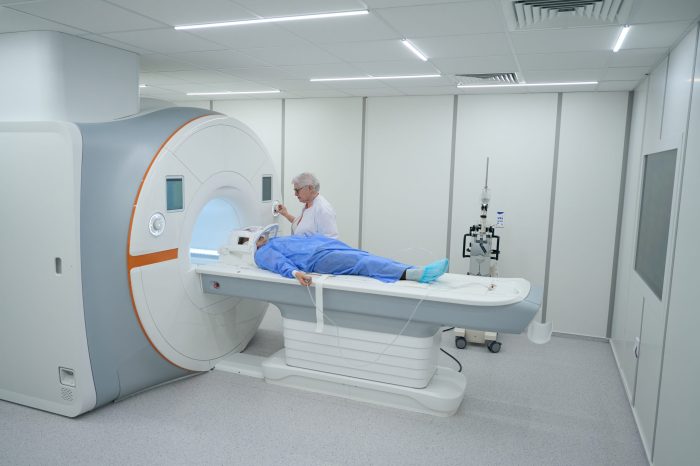
MRI of the knee provides a detailed view of the knee’s intricate structures, offering valuable insights for diagnosis and treatment. This comprehensive guide explores the process, from initial preparation to interpreting results, while highlighting the significance of this imaging technique in evaluating various knee conditions. We’ll delve into the anatomy, procedures, and potential complications, ultimately offering a thorough understanding of MRI of the knee.
This in-depth exploration will cover the different types of MRI sequences used, common indications, and the role of MRI in assessing the severity of knee injuries. We’ll also compare MRI with other imaging modalities, discussing its advantages and disadvantages in various clinical scenarios. Furthermore, the potential complications associated with the procedure will be discussed, along with safety considerations.
Introduction to MRI of the Knee
Magnetic Resonance Imaging (MRI) of the knee is a non-invasive medical imaging technique that uses powerful magnetic fields and radio waves to produce detailed images of the knee joint and surrounding structures. This detailed visualization allows for the assessment of soft tissues, cartilage, ligaments, tendons, and bones, offering crucial insights into potential injuries or abnormalities.The purpose of an MRI of the knee is to provide a comprehensive evaluation of the knee’s anatomy, identifying any structural damage, inflammation, or disease processes that may be causing pain, instability, or other symptoms.
This diagnostic tool aids in accurately diagnosing the underlying cause of knee problems, guiding treatment decisions, and monitoring the progression of conditions. The detailed images provide a more accurate diagnosis than other imaging methods, such as X-rays, which primarily visualize bone structures.
Anatomy Visualized in an MRI of the Knee
An MRI of the knee provides detailed images of various structures within and around the joint. These include the bones of the femur (thigh bone), tibia (shin bone), and patella (kneecap). Crucially, it allows visualization of the soft tissues, including cartilage, ligaments (cruciate ligaments, collateral ligaments), tendons (patellar tendon, quadriceps tendon), menisci (cartilage pads), and bursae (fluid-filled sacs).
The surrounding muscles, fat pads, and joint fluid are also depicted, offering a comprehensive picture of the knee’s complex structure.
Common Indications for Ordering an MRI of the Knee
Various clinical situations warrant an MRI of the knee. Common indications include suspected ligament tears (e.g., anterior cruciate ligament (ACL) tear), meniscus tears, cartilage damage (e.g., osteoarthritis), tendonitis, bursitis, or infections. The presence of chronic knee pain, instability, or swelling, especially when other imaging methods are inconclusive, often leads to an MRI to precisely determine the nature and extent of the pathology.
Furthermore, monitoring the effectiveness of treatments, evaluating the progress of conditions, and ruling out other potential diagnoses are additional reasons for ordering an MRI of the knee.
Possible Knee Conditions and Associated Findings
The following table Artikels various knee conditions, their typical symptoms, potential MRI findings, and general treatment approaches.
| Condition | Symptoms | Possible Findings | Typical Treatment |
|---|---|---|---|
| Meniscus Tear | Pain, swelling, locking, catching sensation in the knee, especially with twisting or pivoting movements. | Irregular or displaced meniscus tissue, focal or diffuse meniscal tears, possible joint effusion (fluid buildup). | Conservative treatment (rest, ice, physical therapy) may be sufficient for minor tears. Surgery (meniscectomy or meniscus repair) may be necessary for significant tears, depending on the location and severity of the tear. |
| Anterior Cruciate Ligament (ACL) Tear | Pain, instability, swelling, a “pop” sound during injury, difficulty bearing weight. | Disruption or complete tear of the ACL fibers, often associated with joint effusion and edema. | Conservative treatment may be suitable for partial tears. Surgical reconstruction of the ACL is frequently required for complete tears, involving grafting and restoring stability. |
| Osteoarthritis | Progressive pain, stiffness, swelling, reduced range of motion, especially in the morning. | Cartilage loss, bone spurs (osteophytes), joint space narrowing, and subchondral bone sclerosis (thickening of the bone under the cartilage). | Pain management, physical therapy, lifestyle modifications, and in severe cases, joint replacement surgery. |
| Patellar Tendinopathy | Pain around the kneecap, especially during activities involving jumping or running. | Thickening or inflammation of the patellar tendon, possible edema or tendon tears. | Conservative treatment, including rest, ice, and physical therapy, focusing on strengthening the surrounding muscles. In severe cases, corticosteroid injections or surgery may be necessary. |
Types of MRI Knee Examinations
MRI of the knee provides valuable insights into the structure and function of the joint. Different MRI sequences are employed to capture various aspects of the knee’s anatomy and pathology, allowing radiologists to assess the extent and nature of any abnormalities. Understanding these sequences is crucial for interpreting the results accurately and guiding appropriate treatment decisions.
Different MRI Sequences
Various MRI sequences are used to obtain detailed images of the knee, each designed to highlight specific tissue characteristics. These sequences differ in their acquisition parameters, enabling the visualization of different aspects of the knee, including bone, cartilage, ligaments, tendons, and menisci.
Proton Density (PD) Weighted Imaging
This sequence provides a relatively balanced representation of the water content within tissues. It’s particularly useful for evaluating the overall morphology of the knee structures and detecting fluid collections or edema. The signal intensity primarily reflects the proton density, thus aiding in differentiating between different tissue types. Advantages include providing a basic anatomical overview and being sensitive to fluid and edema.
Disadvantages are limited ability to distinguish between various tissue types, making it less useful for detailed pathology assessment.
T1-Weighted Imaging
T1-weighted images are highly sensitive to the fat content of tissues. They provide excellent contrast between fat and water, allowing for precise delineation of fat pads, bone marrow, and other fatty structures. This helps in differentiating between fat and other soft tissues. Advantages include excellent visualization of fat and distinguishing fat from water. Disadvantages are that it doesn’t offer a clear representation of fluid and edema.
T2-Weighted Imaging
T2-weighted images are highly sensitive to water content. They excel at depicting edema, fluid collections, and abnormalities within soft tissues. The longer echo time enhances the contrast between fluid and other soft tissues. Advantages include excellent visualization of fluid, edema, and soft tissue abnormalities. Disadvantages include lower resolution for bone and cartilage, and potential for increased noise and image artifacts.
Fat-Suppressed T2-Weighted Imaging
Fat-suppressed T2-weighted images are a modified version of T2-weighted images. They eliminate the signal from fat, enhancing the visibility of soft tissue structures and fluid collections. This helps in reducing the interference from fat signals and improving the visualization of subtle abnormalities in the soft tissues. Advantages include minimizing fat signal, improving the visualization of subtle abnormalities, and enhancing the identification of fluid and edema.
Disadvantages are the need for specific technical parameters, which can occasionally affect the overall image quality.
Contrast Agents in Knee MRI
Gadolinium-based contrast agents are occasionally used in knee MRI. These agents enhance the signal intensity of blood vessels and inflamed tissues. This allows for better delineation of vascular structures and the identification of inflammatory processes. Advantages include better visualization of vascular structures and inflammatory processes. Disadvantages include potential allergic reactions and the risk of nephrogenic systemic fibrosis in specific cases.
Comparison of MRI Sequences
| Sequence | Primary Tissue Contrast | Advantages | Disadvantages |
|---|---|---|---|
| Proton Density (PD) | Water content | Basic anatomy, fluid/edema | Limited tissue differentiation |
| T1-weighted | Fat | Excellent fat visualization | Poor fluid/edema visualization |
| T2-weighted | Water content | Excellent fluid/edema visualization | Lower bone/cartilage resolution |
| Fat-suppressed T2 | Water content (no fat) | Improved soft tissue visualization | Potential for technical issues |
Clinical Applications
MRI of the knee provides invaluable diagnostic information for a wide range of knee conditions. It allows visualization of soft tissues, ligaments, tendons, cartilage, and bone structures, enabling precise assessment of the extent and nature of injuries. This detailed imaging helps clinicians accurately diagnose problems, plan appropriate treatment strategies, and monitor the healing process. Accurate diagnosis is crucial for effective patient management and maximizing positive outcomes.
Diagnostic Capabilities
MRI excels at visualizing soft tissue structures, making it exceptionally useful in diagnosing conditions affecting ligaments, tendons, cartilage, and menisci. The detailed images generated by MRI allow for precise identification of tears, sprains, and other pathologies that might be missed by other imaging modalities. This detailed assessment is vital for formulating an accurate diagnosis and treatment plan.
Getting an MRI of the knee can be a bit uncomfortable, but it’s often the best way to see what’s going on inside. Sometimes, people looking for quick weight loss solutions turn to medications, and unfortunately, some weight loss drugs can lead to acne breakouts. weight loss drugs acne can be a surprising side effect, and it’s crucial to discuss all potential consequences with your doctor.
Ultimately, though, an MRI of the knee provides vital information for proper diagnosis and treatment.
Correlation with Patient Symptoms
MRI findings are meticulously correlated with patient symptoms to arrive at a comprehensive understanding of the knee condition. For instance, if a patient presents with pain and instability in the knee, MRI can reveal a ligament tear, providing a clear link between the physical manifestation and the underlying pathology. This correlation is paramount for effective patient management.
Common Knee Pathologies Detected
MRI is a powerful tool in identifying a wide range of common knee pathologies. These include meniscal tears, ligament injuries (cruciate and collateral), cartilage defects, and bone bruises. Accurate identification of these conditions is crucial for prompt and appropriate intervention.
Assessment of Injury Severity
The severity of knee injuries can be accurately assessed using MRI. The degree of ligament or meniscus tear, the extent of cartilage damage, and the presence of bone contusions can be precisely quantified. This assessment enables clinicians to tailor treatment strategies to the specific needs of each patient.
Table of Common Knee Pathologies and Typical MRI Findings
| Pathology | Typical MRI Findings |
|---|---|
| Meniscal Tear (medial or lateral) | Focal discontinuity or irregularity of the meniscus; high signal intensity on T2-weighted images, indicating fluid or edema within the meniscus; possible visualization of a meniscal fragment or flap. |
| Anterior Cruciate Ligament (ACL) Tear | Abnormal signal intensity or discontinuity of the ACL fibers; edema or fluid collection around the ACL; possible visualization of a complete or partial tear. |
| Posterior Cruciate Ligament (PCL) Tear | Similar to ACL tears, but the findings are often localized to the posterior aspect of the knee joint; disruption or abnormal signal intensity of the PCL fibers. |
| Medial Collateral Ligament (MCL) Tear | Disruption or abnormal signal intensity of the MCL fibers; edema or fluid collection around the MCL; possible visualization of a complete or partial tear. |
| Cartilage Defect (e.g., chondromalacia patella) | Focal loss of cartilage thickness or irregularity; abnormal signal intensity within the cartilage, indicative of degeneration or damage; possible visualization of exposed bone. |
Technical Aspects of the Procedure
Getting an MRI of your knee can feel a bit daunting, but understanding the technical aspects can ease your mind. This section delves into the preparation, procedure, safety measures, and positioning involved in a safe and effective knee MRI. Knowing what to expect can help you feel more comfortable and confident throughout the entire process.
Patient Preparation
Thorough preparation is crucial for a successful MRI scan. Patients are typically asked to remove any metal objects, including jewelry, watches, and hair clips, as these can interfere with the magnetic field. Clothing with metal zippers or buttons should also be avoided. Loose, comfortable clothing that is easily removed is recommended. Patients with pacemakers or other implanted medical devices will need to inform the technician, as these devices may be incompatible with the MRI environment.
Some patients may need to fast or abstain from certain medications prior to the exam. These instructions are always provided by the radiologist or technologist and will vary based on individual circumstances.
Steps in Performing the Knee MRI
The MRI process typically begins with the patient being positioned on a specialized table that slides into the MRI machine. The table is designed to accommodate different body types and positions, ensuring a comfortable and secure placement for the scan. A radiofrequency coil is often placed directly over the knee joint to enhance the signal quality of the images.
Radio waves and a strong magnetic field work together to create detailed images of the knee’s internal structures. The entire process is monitored by trained personnel. The technician will explain any necessary steps and provide support and reassurance throughout the exam.
Safety Considerations and Precautions
MRI procedures are generally safe for most patients. However, some precautions are essential. Patients with metal implants, pacemakers, or other implanted medical devices should inform the MRI technician immediately. Individuals with claustrophobia may find the enclosed space of the MRI machine challenging; they can be offered relaxation techniques or medication to help manage this. Pre-existing conditions like severe heart problems or metal in the eyes need to be discussed with the radiologist prior to the scan.
Specific safety guidelines and protocols are in place to mitigate any potential risks and ensure patient safety during the procedure. A careful assessment of the patient’s medical history is paramount.
Patient Positioning During the Scan
Proper positioning is critical for obtaining high-quality images. The patient is typically positioned supine (lying on their back) with the knee extended. The knee joint is often positioned in a way that allows for optimal visualization of the structures of interest. The technician will carefully position the patient, ensuring the knee is aligned correctly within the field of view.
This precise positioning helps to ensure that the images are accurate and informative.
Procedure Steps and Potential Patient Concerns
| Step | Description | Potential Patient Concerns |
|---|---|---|
| 1. Preparation | Patient removes metal objects, receives instructions, and completes necessary questionnaires. | Anxiety about the procedure, concerns about claustrophobia, discomfort from removing jewelry. |
| 2. Positioning | Patient is positioned on the MRI table with the knee in the correct alignment. | Discomfort from the positioning, concerns about movement during the scan. |
| 3. Scan Acquisition | The MRI machine uses radio waves and a strong magnetic field to create detailed images of the knee. | Noise from the machine, feelings of confinement, anxiety during the scan. |
| 4. Image Analysis | Images are reviewed and analyzed by a radiologist to generate a report. | Waiting for the results, concerns about the findings. |
Interpreting MRI Results

MRI of the knee provides detailed images of the soft tissues and structures, allowing radiologists to identify various conditions. Interpreting these results requires a thorough understanding of normal anatomy and the appearance of different pathologies on MRI. The findings can significantly influence treatment decisions, from conservative management to surgical interventions.Interpreting MRI results is a complex process involving careful observation of image details, correlation with patient history, and clinical examination.
Radiologists utilize specialized software and their expertise to analyze the images, identifying potential abnormalities. The goal is to accurately diagnose the underlying cause of the patient’s knee pain or dysfunction.
Common MRI Findings for Various Knee Conditions
A comprehensive analysis of MRI findings requires familiarity with normal knee anatomy and the appearance of common pathologies. Radiologists meticulously examine the different structures within the knee, including the cartilage, ligaments, tendons, and menisci. Changes in the appearance of these structures can indicate injury or disease.
Examples of How MRI Findings Guide Treatment Decisions
MRI findings can significantly guide treatment strategies. For example, a tear in the anterior cruciate ligament (ACL) visualized on MRI would necessitate a discussion between the patient and physician regarding surgical intervention or a course of physical therapy. Similarly, a diagnosis of osteoarthritis, evidenced by significant cartilage loss, might lead to recommendations for pain management, physical therapy, and potential joint replacement.
These treatment options are tailored to the specific findings and the patient’s overall health.
How Radiologists Analyze MRI Images, Mri of the knee
Radiologists employ a systematic approach to analyzing MRI images. They first examine the overall anatomy of the knee, noting any obvious abnormalities. Then, they focus on specific structures, assessing the integrity of the menisci, ligaments, and tendons. They evaluate the presence of edema, which might suggest inflammation or injury. Finally, they look for bone marrow edema, which is a sign of potential bone injury.
Sophisticated software is often used to measure the extent of damage and assist in the interpretation.
Limitations of MRI in Diagnosing Knee Conditions
While MRI is a powerful diagnostic tool, it does have certain limitations. One limitation is that it may not always accurately identify subtle or early-stage injuries. MRI can also be expensive and may not be readily available in all locations. Additionally, the presence of metal implants or other artifacts can affect image quality and potentially obscure critical findings.
The interpretation of MRI findings also relies heavily on the expertise of the radiologist and may not fully account for all factors in a patient’s clinical presentation.
Comparison of Knee Injury Findings on MRI
| Injury | Typical MRI Appearance | Description |
|---|---|---|
| Meniscus Tear | Focal or diffuse signal abnormality within the meniscus, often with a disrupted or irregular meniscal contour. | Damage to the meniscus, a C-shaped cartilage pad in the knee. Can be partial or complete. |
| ACL Tear | Disruption or discontinuity of the ACL fibers, often accompanied by edema and/or hemorrhage in the surrounding tissues. | Tear of the anterior cruciate ligament, a crucial ligament that stabilizes the knee. |
| MCL Tear | Abnormal signal intensity or discontinuity in the medial collateral ligament (MCL), potentially with edema in the surrounding tissues. | Tear of the medial collateral ligament, which provides medial stability to the knee. |
| Patellar Tendonitis | Edema and thickening of the patellar tendon, sometimes with focal or diffuse signal abnormalities. | Inflammation of the patellar tendon, often associated with overuse or repetitive stress. |
| Osteoarthritis | Cartilage loss, bone marrow edema, subchondral sclerosis, and cysts. | Degenerative joint disease characterized by cartilage breakdown and bony changes. |
Comparison with Other Imaging Techniques
Understanding the knee’s intricacies often requires a multi-faceted approach. Different imaging modalities offer varying degrees of information, and choosing the right one is crucial for accurate diagnosis and treatment planning. This section delves into how MRI compares to other common imaging techniques for knee assessments.
Getting an MRI of your knee can be a crucial diagnostic tool, revealing potential issues like cartilage tears or meniscus problems. While focusing on physical remedies is important, did you know that incorporating foods like apples, and specifically, apple pectin, might offer some support for joint health? Exploring the benefits of apple pectin could potentially aid in overall wellness, contributing to a more comfortable recovery process after an MRI of the knee.
the benefits of apple pectin are definitely worth considering as part of a holistic approach to knee health. Ultimately, a balanced diet and the results of the MRI itself will guide any further treatment plan.
Comparison with X-rays
X-rays are valuable for visualizing bone structures. They effectively detect fractures, dislocations, and significant bone abnormalities. However, X-rays provide limited information about soft tissues, such as ligaments, tendons, and cartilage. MRI, in contrast, excels at visualizing these soft tissues, allowing for detailed assessments of injuries and pathologies not readily apparent on X-rays. For example, a patient presenting with knee pain might show a subtle cartilage tear on MRI, but only a fracture on X-ray.
This highlights the crucial role of MRI in evaluating the full extent of knee damage.
Comparison with CT Scans
CT scans offer detailed cross-sectional views of the knee, showcasing bone structures and soft tissues in great detail. While CT scans can detect fractures and bone abnormalities with precision, their ability to visualize soft tissue structures like cartilage is less comprehensive than MRI. MRI, with its superior soft tissue contrast, provides a more complete picture of the knee’s soft tissue elements, making it ideal for assessing ligament tears, meniscal injuries, and other soft tissue pathologies.
Comparison with Ultrasound
Ultrasound is a valuable tool for evaluating the knee, particularly for assessing fluid collections (effusions) and superficial structures. It’s a non-invasive, real-time imaging technique that can be helpful in guiding injections or assessing tendonitis. However, its ability to penetrate deep tissues is limited, making it less suitable for visualizing deep-seated structures like the meniscus or cruciate ligaments. MRI’s superior depth penetration allows for a more comprehensive assessment of the entire knee joint, making it preferred for evaluating complex pathologies.
Situations Favoring MRI
MRI is the preferred imaging modality in several situations. Its superior soft tissue contrast makes it ideal for diagnosing:
- Meniscal tears
- Ligament injuries (ACL, PCL, MCL, LCL)
- Cartilage damage (chondromalacia patellae)
- Tendon tears (patellar tendon, quadriceps tendon)
- Soft tissue tumors
These conditions necessitate the high-resolution soft tissue imaging provided by MRI.
Situations Favoring Other Imaging Techniques
In certain situations, other imaging techniques might be more suitable. For instance:
- X-rays are often the initial imaging study for suspected fractures.
- CT scans are preferred for evaluating complex bone injuries or when precise bone detail is needed.
- Ultrasound is helpful for guiding procedures, assessing fluid collections, and evaluating superficial structures.
The choice of imaging modality depends on the specific clinical question and the suspected pathology.
Summary Table
| Imaging Technique | Strengths | Weaknesses |
|---|---|---|
| X-ray | Excellent for visualizing bone structures, relatively inexpensive | Limited soft tissue visualization, poor contrast for soft tissue pathology |
| CT Scan | Detailed cross-sectional views of bone and soft tissue, good for complex fractures | Less detailed soft tissue visualization compared to MRI, potential radiation exposure |
| Ultrasound | Real-time imaging, non-invasive, helpful for guiding procedures, assessing superficial structures | Limited penetration depth, not ideal for deep-seated structures |
| MRI | Excellent soft tissue contrast, detailed visualization of ligaments, tendons, cartilage, and menisci | Can be more expensive, longer examination time, contraindications for certain patients (e.g., pacemakers) |
Emerging Trends and Future Directions
The field of MRI knee examinations is constantly evolving, driven by technological advancements and the need for more precise and informative diagnostics. This evolution promises to significantly impact both the accuracy of diagnoses and the development of more targeted treatment strategies. This section explores the exciting innovations shaping the future of MRI knee imaging.
Latest Advancements in MRI Technology
MRI technology is continually improving, leading to higher resolution images, faster acquisition times, and greater versatility in image contrast. These advancements allow for a more detailed visualization of the knee’s complex anatomy, including subtle pathologies that might have been missed in the past. Improvements in gradient coil technology, for example, result in sharper images and reduced imaging artifacts, leading to better visualization of cartilage and meniscus tears.
Getting an MRI of the knee can be a bit nerve-wracking, but it’s often crucial for diagnosing issues. While we’re on the topic of medical imaging, did you know that some women might need to consider how certain medications, like combination birth control pills , might interact with the procedure? Ultimately, it’s always best to discuss any concerns with your doctor before undergoing an MRI of the knee.
Moreover, faster acquisition sequences enable more dynamic imaging, allowing for the assessment of joint motion and identifying subtle abnormalities not apparent in static images.
Impact on Diagnosis and Treatment
The increased resolution and speed of MRI examinations directly translate into improved diagnostic accuracy. Physicians can now identify smaller tears, subtle cartilage defects, and early signs of osteoarthritis with greater certainty. This improved diagnostic capability allows for earlier and more precise interventions, potentially slowing disease progression and improving patient outcomes. For instance, early detection of meniscus tears using high-resolution MRI can lead to prompt surgical intervention, preventing further damage and preserving joint function.
Likewise, accurate assessment of cartilage damage allows for tailored treatment plans, such as targeted therapies or surgical procedures, improving long-term outcomes.
Artificial Intelligence in MRI Analysis
Artificial intelligence (AI) is rapidly transforming the way medical images are analyzed. AI-powered algorithms can automatically detect subtle abnormalities in MRI scans, potentially reducing the workload on radiologists and improving diagnostic speed. This automated analysis also increases the consistency of interpretation across different facilities and clinicians, which is particularly valuable in situations where a second opinion is required or when there’s a need to compare findings across multiple patients.
For example, an AI algorithm could be trained to identify subtle signs of cartilage degeneration, enabling earlier diagnosis and more effective intervention.
Potential Future Applications of MRI
MRI technology holds the potential for numerous future applications in knee imaging. One area of interest is the development of functional MRI techniques, which could assess the biomechanics of the knee joint under various loading conditions. Such analyses could provide valuable insights into the causes of knee pain and guide the development of personalized treatment strategies. Further development of advanced contrast agents could lead to better visualization of specific tissue structures within the knee, providing more comprehensive insights into disease processes.
Finally, the integration of MRI with other imaging modalities, like ultrasound or CT, could provide a more comprehensive assessment of the knee, yielding more complete diagnostic information.
Future of MRI Technology in Knee Imaging
The future of MRI technology in knee imaging promises a significant advancement in diagnostic accuracy and precision. As technology continues to evolve, we can anticipate a decrease in scan times, improvements in resolution, and enhanced visualization of subtle anatomical details. This will allow for a more comprehensive understanding of the knee’s intricate structures and pathologies, ultimately leading to more effective and personalized treatment strategies.
Expect the ability to visualize dynamic processes within the knee, providing crucial information about joint function and identifying subtle signs of early disease. The development of new contrast agents and advanced image analysis techniques will further refine diagnostic capabilities and allow for a more detailed and accurate evaluation of knee conditions.
Potential Complications and Risks: Mri Of The Knee

MRI of the knee, while a valuable diagnostic tool, carries potential complications and risks, although they are generally low. Understanding these factors is crucial for informed patient consent and proactive risk mitigation. Proper patient evaluation and adherence to safety protocols are paramount to minimizing any potential adverse effects.
Potential Complications from the Procedure Itself
The MRI procedure itself is generally safe, but some patients may experience discomfort or anxiety during the examination. The confined space of the MRI machine can trigger claustrophobia in susceptible individuals. Proper pre-procedure communication and relaxation techniques can help address these concerns. Furthermore, some individuals might experience minor physical reactions, such as mild nausea or dizziness. These reactions are usually temporary and resolve on their own.
Risks Associated with Contrast Agents
Gadolinium-based contrast agents are sometimes used in knee MRI scans to enhance the visibility of certain structures. These agents can pose risks, particularly for patients with pre-existing kidney conditions. Severe kidney problems are extremely rare but can occur, especially in patients with compromised renal function. Careful evaluation of renal function and blood tests are crucial before administering any contrast agent.
Furthermore, allergic reactions to contrast agents, although uncommon, can range from mild skin rashes to more serious systemic reactions. Pre-existing allergies, especially to iodine-based contrast agents used in other imaging modalities, increase the risk. Proper pre-procedure screening for allergies and renal function is essential.
Mitigating Risks
To minimize potential risks, careful patient evaluation is critical. This includes thorough medical history review, assessment of kidney function, and screening for allergies. The use of contrast agents should be reserved for specific clinical indications and only after careful consideration of the patient’s individual risk profile. Strict adherence to safety protocols and close monitoring during the procedure further reduces the likelihood of adverse events.
Patient education and preparation can also play a vital role in reducing anxiety and discomfort. Staff training on proper contrast administration techniques is essential.
Potential Side Effects
- Claustrophobia: Some individuals experience anxiety or discomfort in confined spaces. Preparation and communication can help manage this concern.
- Mild discomfort or nausea: These reactions are common and generally transient, resolving on their own.
- Allergic reactions (rare): Symptoms can range from mild skin rashes to more severe systemic reactions. Pre-procedure screening and appropriate management protocols are crucial.
- Kidney problems (rare): Gadolinium-based contrast agents can theoretically affect kidney function, particularly in patients with pre-existing kidney disease. Careful evaluation of renal function and strict adherence to contrast agent usage guidelines are necessary.
Last Word
In conclusion, MRI of the knee is a powerful diagnostic tool, offering detailed visualization of the knee’s complex anatomy. Understanding the procedure, potential complications, and comparison with other imaging techniques is crucial for both patients and healthcare professionals. The information presented here provides a foundation for comprehending the role of MRI in diagnosing and managing various knee conditions.
We hope this guide empowers you to make informed decisions about your health and the use of this vital imaging technique.


